Eating suggestions in Advanced Gall Bladder Cancer

•April 8, 2010 • 1 Comment

Snacks and small meals –

  1. Yoghurts or fromage frais
  2. Dried fruit
  3. Stewed or fresh fruit (bananas are high in calories)
  4. Crisps or nuts
  5. Cheese
  6. Instant soups (make them up with milk to boost calories)
  7. Cereal
  8. Milky drinks
  9. Chocolate
  10. Flapjack

Boost your calorie intake

  1. Mash vegetables with milk and add some grated cheese and egg
  2. Porridge is a very nutritious breakfast – add syrup or sugar and cream
  3. Make up instant soups or gravies with milk instead of water
  4. Add cheese to an omelette
  5. Dip cooked soft vegetables in dips like hummus and sour cream for a healthy snack
  6. Make instant coffee, hot chocolate or Horlicks with full fat milk
  7. Mix a milkshake with ice cream, yoghurt and fresh fruit
  8. Dunk your favourite biscuits into tea and coffee
  9. You can get complete meals in a drink. There are a number of well known ones on the market such as – Ensure, Complan (soups and flavoured drinks) and Fresubin. These can be bought from chemists’ shops or you can get them on prescription from your doctor. Some of these come in 200ml cartons (the size of a small fruit juice carton). Keep one on hand and sip it throughout the day. You can really increase your calorie intake by doing this. Ask your dietitian for advice on what to use and what you can get on prescription.
  10. If you need building up, you can increase the protein content of whole milk by adding a couple of tablespoons of dried milk powder per pint. Use it in the same way as ordinary milk for drinking and cooking. You can also buy protein powders and high energy powders and sprinkle these on everyday foods or add to recipes.

Diarrhoea

If you are having problems with diarrhoea after gallbladder surgery, avoid very high fibre foods (for example, whole grain bread or cereals and dried fruit) as these may make things worse. Tell your doctor or nurse. You may need some medicines to control your symptoms. It is worth asking to see a dietitian to plan a diet that suits you better.

Life after Cholecystectomy (Gall Bladder Removal)

•March 13, 2010 • 7 Comments

The main function of gall bladder is to store bile but not many people know that removing the Gall Bladder does not stop the flow of bile directly into an intestine. Few say that life after having the Gall Bladder removed is pretty normal. However at the effect varies from individual to individual.

Post surgery tips

  • Dissolvable stitches will usually disappear in around one to three weeks, and sometimes six weeks, depending on the type of stitches you have.
  • Try to get some advice about caring for healing wounds before you go home with your nurse.
  • You may be given special compression stockings to wear after surgery to decrease the possibility of blood clots forming in your legs.
  • For extra insurance against blood clots, plastic boots or leggings are applied over pressure stockings or ace wraps and connected to an air pump machine. The pump rhythmically tightens and loosens different parts of the boots, helping to push the blood back up to the heart.

Food

  • To be on the safer side it is advisable to start on clear liquids post surgery, then full liquids and finally a regular diet.
  • Should avoid too greasy and too spicy food.
  • Food with red or black pepper may cause discomfort.
  • Whole grain cereals and breads, fresh fruit and fresh vegetables should be included in his daily menu.
  • Regular bowel movements can be difficult after surgery. Don’t strain if the stool is too hard. Walking will help to stimulate the bowels.
  • Eating foods rich in fiber, such as fruit, bran, cereal, and beans, will also help restore regularity.
  • Drink plenty of liquids; prune juice may help make the stool softer. Or, if your doctor approves, you can take an over-the-counter fiber laxative.

Possible complications

  • Pain in your abdomen, bloating, wind and Diarrhea
  • Acid Reflux, Nausea, Stomach Cramps and Spasm, Fullness, Burning in stomach
  • Blockage of a blood vessel with a bubble of carbon dioxide gas may occur.
  • In some cases during surgery Dr pumps small amount of air into tummy. They cannot remove all the air so some is left behind to come out on its own. The left behind air causes pain in the shoulders as well.

Call Doctor If…

  • Pain gets worse.
  • Your incision is swollen and red, or you see any pus. These are signs of infection.
  • Your stitches or staples come apart.
  • Your bandage becomes soaked with blood.
  • You develop a high temperature.
  • You see any of the following signs that too much bile is building up in your body: Yellow skin, Light brown or yellow stool, Dark yellow or light brown urine.

Seek Care Immediately If…

  • You develop chest pain or sudden trouble breathing

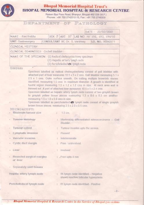
Post surgery biopsy report of my mother

•March 13, 2010 • 1 Comment
Biopsy Report

Biopsy Report

My mother has recently gone thru the stage IV GBC surgery of 16-Feb 2010. She is at home now and would be going through post surgery Radiotherapy next week. I have her post surgery biopsy reports and wanted to share them over here for comments.

GBC is a very rare disease and it would be great if someone can suggest anything relevant.

Chemotherapy

•September 18, 2009 • 1 Comment

Chemotherapy is a drug treatment that uses powerful chemicals to kill fast-growing cells in your body. Chemotherapy is most often used to treat cancer, since cancer cells grow and multiply much more quickly than most cells in the body.

Many different chemotherapy drugs are available and alternative approaches to chemotherapy may help make treatments more powerful while limiting side effects.

Chemotherapy resistance testing. Before your treatment begins, your doctors can perform laboratory tests on tumor cells to determine which drugs are likely to be most effective against the cancer. Not only does testing limit unnecessary exposure to potentially toxic chemotherapy agents, but it can be especially valuable when there’s a choice between two or more possible treatments.

Metronomic chemotherapy. The chemotherapy is delivered in small, regular amounts over a prolonged period rather than all at once. While the individual doses used in metronomic chemotherapy are smaller than typical doses, giving them at frequent intervals can not only shrink the tumor by inhibiting the growth of the blood vessels that feed it, but may also destroy cancer cells.This approach is also less toxic and easier to tolerate than traditional chemotherapy, in which the maximum tolerated dose (MTD) is used.

Intra-arterial chemotherapy (IAC). This treatment is used most commonly in the treatment of liver cancer, but may also be used for head, face, neck, pancreatic, and pelvic cancers. The medication is delivered through the arteries that lead to the site of the tumor. The goal is to subject the cancer to a very high dose of chemotherapy while delivering less to the rest of your body.

Intraperitoneal chemotherapy. In this treatment for ovarian cancer, chemotherapy is administered directly to your peritoneal area, or abdominal cavity, to prevent tumor cells from forming.

Chemotherapy drugs can be used alone or in combination to treat a wide variety of cancers. While traditional chemotherapy agents aim to destroy rapidly dividing cells, new types of drugs interfere with the development of cancer at the molecular level. Because these drugs target specific molecules, they’re sometimes called molecular-targeted treatments.

Targeted therapies are more selective than traditional cancer therapies and spare more healthy cells. Ultimately researchers hope to develop targeted therapies tailored to the unique molecular characteristics of each individual’s tumor, resulting in truly personalized treatment. Many targeted therapies are still in development, but others are in clinical trials or have already been approved by the FDA. They are being tested for use alone, in combination with other targeted therapies, and with conventional treatments, such as traditional chemotherapy.

Anti-angiogenesis drugs: Chemicals in the body control the process of angiogenesis, or the formation of new blood vessels. These drugs are designed to stop the spread of cancer by cutting off the blood supply that feeds tumors. Several anti-angiogenesis medications are already available and are used in coordination with standard chemotherapy to increase its effectiveness.

Apoptosis-inducing drugs: Also known as programmed cell death (PCD), apoptosis is the orderly process of cell death by which the body disposes of abnormal, diseased, or unnecessary cells. Unlike normal cells, however, cancer cells are unable to undergo apoptosis. These drugs stimulate cancer cell death by interfering with proteins and other substances in tumor cells that cause them to live longer than normal cells.

Differentiation drugs: These drugs cause immature cells to become more differentiated, making them function more like normal cells and limiting the uncontrolled growth associated with cancer. This approach has potential for cancer prevention, and the FDA has already approved one such drug to protect people who have a high risk of developing colon cancer.

Signal transduction inhibitors: These drugs prohibit the spread of cancer cells by interfering with the communication signals they rely on to grow. Also present naturally in foods such as soy and citrus fruits, signal transduction inhibitors may help stop cancer from spreading to healthy tissue.

Though chemotherapy is an effective way to treat many types of cancer, chemotherapy treatment also carries a risk of side effects. Some chemotherapy side effects are mild and treatable, others can cause serious complications.

CA 19-9

•September 16, 2009 • 7 Comments

Formal name: Cancer Antigen 19-9

Cancer antigen 19-9 (CA 19-9) is a protein that exists on the surface of certain cells. CA 19-9 does not cause cancer; rather, it is a protein that is shed by the tumor cells, making it useful as a tumor marker to follow the course of the cancer.

CA 19-9 is elevated in most patients with advanced pancreatic cancer, but it may also be elevated in other cancers, conditions, and diseases such as colorectal cancer, lung cancer, gall bladder cancer, gall stones, pancreatitis, cystic fibrosis, and liver disease. Other causes of bile duct obstruction may also cause very high CA 19-9 levels, which fall when the blockage is cleared. It is often a good idea, if the bile ducts are blocked, to wait a week or two after the blockage is removed or treated to check CA 19-9 levels. If they are checked inititally, then it is a good idea to repeat the test after the blockage is removed or treated to see if the cause of the increased CA 19-9 was the tumor or the blockage itself. Very small amounts of CA 19-9 may also be found in healthy patients.

Related Tests: Bilirubin, CEA, Liver panel, Tumor markers

Why Get Tested?

To help differentiate between cancer of the pancreas and bile ducts and other conditions; to monitor response to pancreatic cancer treatment and to watch for recurrence

When to Get Tested?

When your doctor suspects that you have pancreatic cancer and during or following pancreatic cancer treatment

How is the sample collected for testing?

A blood sample is obtained by inserting a needle into a vein in the arm.

How is it used?

CA 19-9 is not sensitive or specific enough to be considered useful as a tool for cancer screening. Its main use is as a tumor marker: 

  • to help differentiate between cancer of the pancreas and bile ducts and other non-cancerous conditions, such as pancreatitis; 
  • to monitor a patient’s response to pancreatic cancer treatment; and
  • to watch for pancreatic cancer recurrence.

CA 19-9 can only be used as a marker if the cancer is producing elevated amounts of it; if CA 19-9 is not initially elevated, then it usually cannot be used later as a marker.

When is it ordered?

CA 19-9 may be ordered along with other tests, such as carcinoembryonic antigen (CEA), bilirubin, and/or a liver panel, when a patient has symptoms that may indicate pancreatic cancer, including abdominal pain, nausea, weight loss, and jaundice.

If CA 19-9 is initially elevated in pancreatic cancer, then it may be ordered several times during cancer treatment to monitor response and, on a regular basis following treatment, to help detect recurrence.

What does the test result mean?

Low amounts of CA 19-9 can be detected in a certain percentage of healthy people, and many conditions that affect the liver or pancreas can cause temporary elevations.

Moderate to high levels are found in pancreatic cancer, other cancers, and in several other diseases and conditions. The highest levels of CA 19-9 are seen in excretory ductal pancreatic cancer — cancer that is found in the pancreas tissues that produce food-digesting enzymes and in the ducts that carry those enzymes into the small intestine. This tissue is where 95% of pancreatic cancers are found.

Serial measurements of CA 19-9 may be useful during and following treatment because rising or falling levels may give your doctor important information about whether the treatment is working, whether all of the cancer was removed successfully during surgery, and whether the cancer is likely returning.

Is there anything else I should know?

Unfortunately, early pancreatic cancer gives few warnings. By the time a patient has symptoms and significantly elevated levels of CA 19-9, their pancreatic cancer is usually at an advanced stage.

Why is my doctor not screening me for CA 19-9?

CA 19-9 is not sensitive or specific enough to be recommended as a screen for people who do not have symptoms. There are too many false positives and false negatives associated with it. Researchers are searching for other markers that may help detect pancreatic cancer at an earlier stage and that may be more suitable for screening.

What other procedures will my doctor likely order along with my CA 19-9?

Your doctor may order a CT scan (computed tomography), an ultrasound, an MRCP (using an MRI scan to look at the pancreatic and bile ducts), an ERCP (endoscopic retrograde cholangiopancreatography, a procedure in which a small lighted tube is passed through the mouth and stomach into the small intestine and then into the bile and pancreatic ducts), and/or a biopsy to look for cancer cells under the microscope.

What are the main risk factors for pancreatic cancer?

Doctors still do not know what causes most cases of pancreatic cancer. Identified risk factors include smoking, age (most are over 50 years old), gender (males are more likely to have it than females), family history, diabetes, chronic pancreatitis, and heavy occupational exposure to certain chemicals and dyes.

Tumors Hungry For Sugar: Findings Point To New Ways To Fight Cancer

•August 27, 2009 • Leave a Comment

McClatchy-Tribune Information Services — Unrestricted

August 22, 2009

University of Utah biochemists have made a breakthrough in understanding how cancer cells feed on glucose, possibly paving the way for new drugs designed to starve cancer into submission.

Cancer cells use glucose in tandem with another crucial nutrient, the protein glutamine, an amino acid found in many foods, according to findings published this week by researchers at the Huntsman Cancer Institute. The findings could spur development of new chemotherapies that would stall tumor growth by deactivating cancer cells’ ability to use glucose, said Don Ayer, a professor of oncological sciences whose lab published the research in the Proceedings of the National Academy of Science .

For decades, science has known that cancer cells suck up inordinate quantities of glucose, nature’s ubiquitous biological fuel, in a process that quickly blows tiny tumors into deadly malignancies.

PET scans use cancer cells’ high rate of glucose metabolism to build images of tumors. These cells also need glutamine, just like normal cells.

“It’s absolutely clear you need both for tumor growth. They seem to need it more than other nutrients. If you deprive them of one or the other, tumors don’t grow,” Ayer said.

Mohan Kaadigea, a postdoctoral researcher in Ayer’s lab, spearheaded the study, whose co-authors include Ayer; Sadhaasivam Kamalanaadhana, also a member of the Ayer lab; and Ryan Looper, an assistant professor in the Department of Chemistry.

The lab’s work, funded by the National Institutes of Health and the American Cancer Society, seeks to unlock the molecular mysteries associated with tumor proliferation.

“Research into the factors that regulate the metabolism and growth of cancer cells is still at an early stage,” said Janet Shaw, a U. professor in the Department of Biochemistry and a former Huntsman researcher. “Dr. Ayer’s discovery that glutamine and glucose utilization are linked is important because it identifies a number of new molecular targets that could be manipulated to interfere with the growth and survival of tumor cells.”

This week’s discovery builds on the lab’s previous research identifying the role of MondoA, a protein that switches genes on and off, in tumorigenesis. This protein affects the gene TXNIP, which suppresses tumor growth by blocking glucose uptake into cancer cells. The Ayer team discovered that in the presence of glutamine, MondoA deactivates TXNIP. This is important because it suggests new ways to impede tumor growth.

“If you don’t have glutamine, the cell is short-circuited due to a lack of glucose, which halts the growth of the tumor cell,” Ayer said.

The next step is to learn how the Mondo protein works in relationship with glutamine.

“If you can modify the metabolism of the tumor cell you can have a benefit. This is not a new idea,” Ayer said. “If we can figure out how glutamine signals to Mondo, that has quite a bit of chemotherapeutic potential.”

Were it developed, a drug that blocks glucose uptake would not likely choke off normal cell growth, as many cancer chemotherapy drugs currently do because of their toxicity.

“Tumor cells seem to be addicted to glucose. Normal cells are not. They grow at a slower rate and if you challenge them with nutrient deprivation they can be more flexible,” Ayer said.

Ayer emphasized that his lab’s findings shed no light on dietary impacts on tumor growth. Glutamine is the most common amino acid in our bodies and glucose levels are tightly regulated by our endocrine system, regardless of sugar consumption.

Cancer and diet

The fact that cancer cells might die if deprived of glucose doesn’t mean cancer patients should cut sugar out of their diets, researchers say.

Cancer patients should eat a balanced diet to promote good health. Cutting out sugar would not inhibit tumor growth, said Don Ayer, a professor of oncological sciences at the Huntsman Cancer Institute. Even with a sugar-free diet, there still would be plenty of glucose in the blood to feed cancer.

Copyright (C) 2009, The Salt Lake Tribune

Causes & risk factors for gallbladder cancer

•August 12, 2009 • 1 Comment

Researchers have found several risk factors that make a person more likely to develop gallbladder cancer.

Most doctors studying the subject think that chronic inflammation is the major cause of gallbladder cancer. When the gallbladder release bile slowly, the gallbladder tissue is exposed to the bile for longer than usual. This may lead to irritation and inflammation. Scientists also suspect that this longer exposure to possible cancer-causing substances in the bile could also be responsible. Certain abnormalities in the ducts that carry fluids from the gallbladder and pancreas to the small intestine can cause juices from the pancreas to flow backward into the gallbladder and bile ducts. Researchers suspect that this reflux (backward flow) of pancreatic juices may irritate the cells lining the gallbladder and bile ducts in a way that causes irritation and inflammation. This may stimulate their growth and perhaps make them more sensitive to cancer-causing substances.

Risk factors for gallbladder cancer 

  • Gallstones: Gallstones are the most common risk factor for gallbladder cancer.
  • Porcelain gallbladder: Porcelain gallbladder is a condition in which the wall of the gallbladder becomes covered with calcium deposits.
  • Female gender: In the United States, gallbladder cancer occurs more than twice as often in women. Gallstones and gallbladder inflammation, 2 important risk factors for gallbladder cancer, are much more common among women than men.
  • Obesity: Patients with gallbladder cancer are more often overweight or obese than people without this disease.
  • Older age: While it can occur at younger ages, gallbladder cancer is seen mainly in older people. The average age at the time of diagnosis is 73. Almost 3 out of 4 people with gallbladder cancer are older than age 65 when it is found.
  • Ethnicity: Native Americans, particularly in the southwestern United States, and Mexican Americans have a higher rate of gallbladder cancer.
  • Choledochal cysts: Choledochal cysts are bile-filled sacs that are connected to the common bile duct, the tube that carries bile from the liver and gallbladder to the small intestine. The cysts can grow over time and may contain as much as 1 to 2 quarts of bile. The cells lining the sac often have areas of pre-cancerous changes, which increase a person’s risk for developing gallbladder cancer.
  • Abnormalities of the bile ducts: The pancreas is another organ that releases fluids through a duct into the small intestine to aid digestion. This duct normally meets up with the common bile duct just as it enters the small intestine. Some people have abnormalities where these ducts meet that allow juice from the pancreas to reflux (flow back “upstream”) into the bile ducts. This backward flow also prevents the bile from being emptied through the bile ducts as quickly as normal. These people are at higher risk of gallbladder cancer.
  • Gallbladder polyps: A gallbladder polyp is a growth that bulges outward from the surface level of the inner gallbladder wall. Some polyps are formed by cholesterol deposits in the gallbladder wall. Others may be small tumors (either cancerous or benign) or may be caused by inflammation. Polyps larger than 1 centimeter (a little less than half an inch) are more likely to be malignant, so doctors often advise removing the gallbladder in patients with gallbladder polyps that size or larger.
  • Industrial and environmental chemicals: It is not clear if exposure to certain chemicals in the workplace or the environment increases the risk of gallbladder cancer. This is a difficult area to study because this cancer is not common. Some animal studies have suggested that chemical compounds called nitrosamines may increase the risk of gallbladder cancer. Other studies have found that workers in the rubber and textile industries may have more gallbladder cancers than the general public.
  • Typhoid: People chronically infected with salmonella (the bacterium that causes typhoid) and those who are carriers of the disease are more likely to develop gallbladder cancer than
    those not infected. Typhoid is rare in the United States.
  • Family history: A history of gallbladder cancer in the family seems to increase a person’s chances of developing this cancer, but the risk is still low because this is a rare disease.

Diagnosis of Gallbladder Cancer

•August 12, 2009 • Leave a Comment

Only about 1 out of 3 gallbladder cancers are found before they have spread to other tissues and organs. Many of these early cancers are found unexpectedly when a person’s gallbladder is removed for treatment of gallstones or chronic (long-term) gallbladder inflammation.

Gallbladders removed for those reasons are always looked at under a microscope by a pathologist (a doctor specializing in lab tests) to see if they contain cancerous cells.

What is gallbladder cancer and types of gallbladder cancers?

•August 12, 2009 • Leave a Comment

Gallbladder cancer is a cancer that starts in the gallbladder.

More than 9 out of 10 gallbladder cancers are adenocarcinomas. An adenocarcinoma is a cancer that starts in cells with gland-like properties that line many internal and external surfaces of the body.

Papillary adenocarcinoma (or just papillary cancer) – These are gallbladder cancers whose cells are arranged in finger-like projections when viewed under a microscope. In general, papillary cancers are not as likely to invade the liver or nearby lymph nodes. They tend to have a better prognosis than most other kinds of gallbladder adenocarcinomas.

About 6% of all gallbladder cancers are papillary adenocarcinomas. There are other types of cancer that can develop in the gallbladder, such as adenosquamous carcinomas, squamous cell carcinomas, and small cell carcinomas, but these are uncommon.

What is gallbladder?

•August 12, 2009 • Leave a Comment

The gallbladder is a small, pear-shaped organ located under the right lobe of the liver. Both the liver and the gallbladder are behind the right lower ribs. The gallbladder is usually about 3 to 4 inches long and normally no wider than 1 inch. intestineThe gallbladder concentrates and stores bile, a fluid made in the liver. Bile helps digest the fats in foods as they pass through the small intestine. Bile may be released from the liver directly into the small intestine, or it may be stored in the gallbladder and released later. When food (especially fatty food) is being digested, the gallbladder contracts and releases bile through a small tube called the cystic duct. The cystic duct joins up with the hepatic duct, which comes from the liver, to form the common bile duct. The common bile duct empties into the small intestine.

The gallbladder is helpful, but it is not essential for life. Many people live normal lives after having their gallbladders removed.